



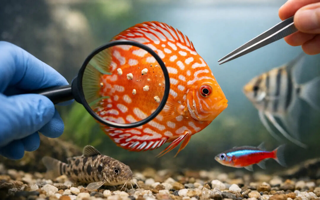
A fish that flashes once on decor may be irritated. A fish that flashes repeatedly, clamps its fins, breathes harder, and develops excess mucus is telling you something more specific. If you want to know how to identify fish parasites, the first rule is simple – do not start with the medicine bottle. Start with observation.
Parasites are often blamed for every spot, scrape, and breathing problem in ornamental fish. That is where many losses begin. White dots are not always ich. Rapid breathing is not always gill flukes. A fish rubbing against the bottom may have skin irritation from water quality, bacterial disease, or a true parasitic infestation. Correct diagnosis matters because treatment success depends on matching the parasite, the host species, and the stage of the disease.
How to identify fish parasites by pattern, not guesswork
The most reliable approach is to read the whole pattern of disease rather than one isolated symptom. Start with three questions. Where on the fish do you see the main change – skin, fins, gills, eyes, or abdomen? How is the fish behaving – flashing, isolating, darting, hanging near the surface, losing appetite, or breathing rapidly? And how fast is the problem spreading through the system?
External parasites usually produce visible and behavioral clues before they kill. Fish may scratch against hard surfaces, produce a gray or bluish mucus film, clamp fins, or show small hemorrhages and erosions. Gill parasites often show up first as respiratory distress. The fish may pump its opercula rapidly, stay near strong aeration, or breathe with one gill more open than the other. Internal parasites are more difficult. You may see weight loss despite feeding, white stringy feces, abdominal swelling, darkened body color, or chronic weakness.
This is why experienced keepers do not diagnose from a single photo if the fish cannot be examined closely. A good diagnosis combines what you see on the body with what the fish is doing and how the group is responding.
Start with the fish’s behavior
Behavior is often the earliest practical clue. Flashing and rubbing suggest irritation of the skin or gills. That irritation may come from protozoan parasites such as Ichthyophthirius, Costia, Chilodonella, or Trichodina, but also from flukes and poor water conditions. The difference is usually in the accompanying signs.
A fish with ich often continues to feed in the early phase while showing repeated flashing and small white cyst-like spots. A fish with Costia may become dull, clamp its fins, stop feeding earlier, and develop a cloudy skin appearance instead of distinct white grains. Chilodonella often produces heavy mucus and respiratory stress, especially in weakened fish or in cooler systems. Trichodina may cause irritation and excess mucus but can be less dramatic until the burden becomes high.
Watch the gills closely. Gill movement that is faster than normal, especially in the absence of obvious ammonia or oxygen problems, should always raise suspicion. Flukes and microscopic protozoa can damage gill tissue quickly. A fish that eats poorly, separates from the group, and breathes hard is rarely dealing with a harmless issue.
Read the skin and fins carefully
Visible changes on the skin are useful, but only if you interpret them carefully. Distinct white spots the size of salt grains point toward ich, especially if they appear on fins and body together. But many keepers mistake air bubbles, debris, epithelial reactions, or healing tissue for parasite spots.
A velvet-like dusting, especially under angled light, may suggest Amyloodinium in marine fish or velvet-type infections in freshwater systems. This appearance is finer than ich and often accompanied by severe respiratory distress. Fish may hold their fins tightly to the body and avoid bright light.
A gray, bluish, or opaque film over parts of the skin often suggests excess mucus production. That response is common with Costia, Chilodonella, and some fluke infestations. You may also see frayed fins, red patches, or ulcers, but here caution is essential. Parasites often open the door to secondary bacterial infection, and then the fish shows a mixed picture. At that point, the parasite may be the trigger, but not the only problem that needs treatment.
Larger visible parasites can sometimes be seen without magnification. Fish lice, anchor worms, and some leeches are macroscopic enough to identify directly. They do not look like dust or tiny grains. They look like attached organisms or protruding structures. These cases are easier to recognize, but even then, secondary inflammation around the attachment site can confuse the picture.
How to identify fish parasites in the gills
Gill disease is one of the most urgent forms of parasitic infection because fish can die before external signs become obvious. When learning how to identify fish parasites, many keepers focus on body spots and miss the gills entirely.
A fish with gill parasites may show rapid breathing, flared opercula, hanging in current, or staying near the water surface. Sometimes one gill cover remains more closed because the fish is protecting damaged tissue on that side. In severe cases the gill tissue becomes pale, swollen, or covered with excess mucus. You may not see this clearly unless the fish is examined in good light.
Gill flukes are a classic cause, but not the only one. Ich, velvet, Costia, and Chilodonella can all affect the gills. Water quality problems can look similar, which is why respiratory distress must always be interpreted alongside water testing and the rest of the clinical picture. If several fish begin breathing heavily after a maintenance error, suspect environment first. If one species declines while others seem normal and flashing has been present for days, parasites move higher on the list.
Internal parasites need a different mindset
Not every parasitic problem is on the skin. Internal flagellates, worms, and other intestinal parasites often produce slower, less dramatic signs. The fish may continue eating but lose weight, develop a pinched abdomen, or pass pale stringy feces. In cichlids and some marine species, chronic wasting with reduced activity often leads keepers to suspect internal parasites, but that suspicion still needs care. Similar signs can occur with malnutrition, chronic bacterial disease, organ failure, or social stress.
Here, time course matters. A fish that has slowly become thin over weeks while others remain normal suggests an individual internal issue. A group that suddenly stops eating and darkens in color suggests something more acute. Internal parasitism is real, but it is also overdiagnosed when the actual cause is feeding imbalance or long-term husbandry stress.
The microscope changes everything
If you are serious about diagnosis, skin scrapes and gill biopsies examined under a microscope are the fastest way to stop guessing. This is where practical fish health management separates itself from trial-and-error treatment.
Many important parasites are microscopic. Costia is tiny and easy to miss without experience. Chilodonella has a characteristic gliding motion. Trichodina appears as a rotating disc. Flukes are larger and often easier to recognize, and ich has a distinctive ciliated body. Once you have seen these organisms clearly, treatment decisions become much more accurate.
This is also why visual documentation is so valuable. Good diagnostic references with representative color photographs and microscopy images train your eye to connect external symptoms with the organism behind them. Gerald Bassleer Books has built much of its practical value around exactly this step – helping keepers move from symptom to verified diagnosis instead of treating blindly.
Common mistakes when identifying parasites
The biggest mistake is treating every white mark as ich. The second is assuming every flashing fish has parasites. The third is ignoring mixed infections.
Fish often present with more than one problem at the same time. A parasite may damage the skin first, then bacteria invade, and finally water quality worsens because feeding and filtration are disrupted. If you only chase the first visible sign, you may miss the condition now causing the real danger.
Another common error is diagnosing too late. By the time ulcers, heavy slime, and collapse are obvious, the parasite burden may already be severe. Earlier clues are subtler – one fish hanging back, light flashing, reduced appetite, slightly faster gill movement. Advanced keepers learn to respect those small changes.
Quarantine history also matters. Fish that have been recently imported, moved, mixed with new stock, or stressed by transport are more likely to show parasitic outbreaks. That does not prove parasites, but it raises the index of suspicion.
What to do before you treat
Observe the affected fish closely, test the water, compare symptoms across the group, and if possible confirm with microscopy. Take clear photos under neutral light. Note whether lesions are raised, flat, dusty, patchy, or ulcerated. Watch feeding response and count gill movements if breathing seems abnormal.
Then decide how confident the diagnosis really is. If you are unsure whether the issue is ich, velvet, flukes, or mucus-producing protozoa, broad assumptions can lead to the wrong treatment or a treatment that is too weak for the real pathogen. This is especially risky in marine systems, with sensitive species, and with invertebrates in the tank.
The best fish keepers are not the ones who medicate fastest. They are the ones who observe best, confirm when possible, and recognize when one symptom can have several causes.
When a fish looks unwell, precision saves lives. The more carefully you learn to read behavior, skin, gills, and progression, the less often parasites will surprise you – and the more often your treatment will actually work.
Recent Comments