



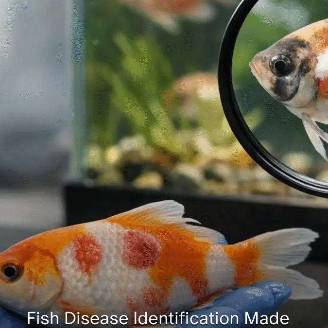
A fish that isolates near a filter outlet, breathes rapidly, and darkens in color has not yet been diagnosed. It has presented a clinical picture. Fish disease identification begins when the keeper resists the urge to name a disease from one visible sign and instead asks what the whole picture is saying.
That distinction prevents costly mistakes. White spots are not always the same parasite. Red skin can indicate injury, poor water quality, bacterial involvement, or parasite irritation. Loss of appetite can occur in nearly every serious health problem, but it can also follow transport, social stress, or a sudden temperature change. Accurate diagnosis is a process of observation, exclusion, and confirmation whenever possible.
Fish Disease Identification Starts With a Case History
Before inspecting the fish closely, establish what changed. The history often reveals more than the most obvious symptom. Record the species affected, the number of fish involved, when signs first appeared, and how quickly the problem progressed. A single newly purchased fish with frayed fins represents a different risk pattern than several established fish becoming lethargic within 24 hours.
Ask practical questions. Were new fish, plants, live foods, or equipment introduced? Has there been a recent power interruption, filter cleaning, medication course, handling event, spawning episode, or change in feed? Are only one species, one size class, or one tank affected? In ponds and aquaculture systems, consider recent weather changes, stocking density, harvesting, and fluctuations in oxygen availability.
The time course matters. Acute disease can develop over hours or days and is often associated with water-quality failures, toxic exposure, oxygen depletion, or rapidly progressing infections. Chronic disease develops over weeks or months and may point toward nutrition, parasites, persistent environmental stress, or an underlying management issue. This does not establish a diagnosis by itself, but it narrows the field of reasonable possibilities.
Examine the Environment Before Treating the Fish
Fish live inside their environment. When water conditions are wrong, the environment may be the primary cause of illness, the trigger that allows infection to take hold, or the reason a treatment fails. Testing water is therefore not a separate chore from diagnosis. It is part of the examination.
Measure temperature, dissolved oxygen when possible, pH, ammonia, nitrite, and nitrate. In marine systems, verify salinity and alkalinity. In recirculating aquaculture systems, review biofilter performance, flow, solids management, and recent feeding rates. Test results should be interpreted alongside the species, system, and recent history. A value that is tolerable for one fish may be harmful to another, particularly when combined with low oxygen or crowding.
Do not dismiss a water-quality problem because the water looks clear. Ammonia and nitrite are invisible. Low oxygen may be most severe before dawn. A filter that appears to be running can still be biologically impaired after overcleaning, medication, or a sudden increase in organic waste.
Observe fish behavior before disturbing the tank or pond. Gasping at the surface, gathering at inflow points, flashing against objects, clamped fins, erratic swimming, loss of equilibrium, and isolation all provide diagnostic information. Note whether normal feeding behavior has changed. In many species, reduced feeding is an early warning sign that appears before clear external lesions.
Build the Clinical Picture Systematically
A disciplined external examination moves from general to specific. First look at the group. Then look at individual fish. Compare affected fish with healthy fish of the same species, age, and sex whenever possible.
Start with body shape and condition. Is the fish thin despite eating? Is the abdomen swollen? Are the eyes sunken or protruding? Is the back unusually curved? Then inspect the skin, scales, fins, gills, mouth, and eyes. Good lighting and a clear observation container can make subtle signs visible without excessive handling.
Skin, scales, and fins
White grains resembling salt may suggest ich, but irregular white patches can also be excess mucus, fungal-like growth, epithelial damage, or another parasite. A gray or blue film on the skin may indicate heavy mucus production, often a response to irritation. Ulcers, hemorrhages, and missing scales require careful interpretation because aggression, net damage, and bacterial disease can look similar at first glance.
Fin erosion is equally nonspecific. It may result from poor water conditions, biting by tank mates, mechanical damage, or bacterial infection. Look at the edge of the fin, the speed of progression, and whether several fish are affected. A clean split in a fin is different from a rapidly receding, inflamed margin.
Gills and breathing
Respiratory distress deserves immediate attention. Rapid opercular movement, gasping, hanging at the surface, or gathering near aeration may indicate low oxygen, ammonia or nitrite exposure, gill parasites, gill infection, or damage from inappropriate chemicals. If several fish show respiratory signs at once, test environmental conditions first and increase aeration while the investigation continues.
Gill color can be informative when examination is safe and justified. Healthy gills are generally red to pink, depending on species. Pale gills can be associated with anemia, while dark, swollen, patchy, or mucus-covered gills may support other concerns. However, gill examination is invasive and should not become casual handling. For valuable stock or repeated losses, professional diagnostic support and laboratory examination may be the most responsible choice.
Feces, appetite, and body condition
Long, pale feces are often blamed on internal parasites, but they are not proof of one. Fish that have stopped eating may pass little material or mucus-like feces regardless of the original cause. A better assessment combines appetite, weight loss, abdominal shape, population history, and, where appropriate, fecal or microscopic examination.
A fish that eats eagerly but loses condition raises different questions than a fish that refuses feed after a temperature drop. Feeding behavior should be documented, not treated as a simple yes-or-no observation.
Use Differential Diagnosis, Not Symptom Matching
The most common diagnostic error is treating a symptom as though it were a disease name. A better method is differential diagnosis: list the plausible causes, then seek observations or tests that separate them.
For example, flashing may be caused by external parasites, poor water quality, irritating chemicals, or social conflict. If flashing begins after a water change and multiple fish show rapid breathing, water chemistry or contamination moves higher on the list. If new fish were introduced and the behavior slowly spreads, parasites become more plausible. A skin scrape examined under a microscope can provide confirmation that observation alone cannot.
Microscopy is one of the most valuable practical skills in fish health. Skin and gill scrapes, performed correctly and humanely, can reveal many external parasites, excess mucus, and some fungal or bacterial patterns. Microscopy does not replace full diagnostic work, and it has limits. Internal disease, viruses, nutritional problems, and many bacterial infections cannot be reliably diagnosed from a simple wet mount. Still, it transforms many cases from guesswork into evidence-based decisions.
Treat the Cause and Protect the Population
When disease is suspected, the first response should stabilize the system. Correct water-quality failures, improve oxygenation, remove obvious stressors, and stop unnecessary handling. Isolate affected fish when practical, but remember that isolation is not always possible or beneficial in a large system where exposure has already occurred. A hospital tank is useful only if its water quality and temperature can be maintained safely.
Avoid stacking medications because symptoms are alarming. Combining products can create additional stress, reduce oxygen, impair filtration, or obscure the diagnostic picture. Medication should follow a working diagnosis, a review of species sensitivity, and a clear plan for dose, duration, water conditions, and follow-up. In food-fish production, legal use requirements and withdrawal periods must be followed without exception.
Keep a written case record. Include water results, dates, mortality, feeding response, treatments, photographs, and microscopic findings. Over time, these records reveal patterns that memory misses. They also make expert consultation far more efficient because the case can be evaluated from evidence rather than fragments of recollection.
Know When the Case Needs Escalation
Some situations justify immediate professional assistance: unexplained mortality, severe respiratory distress, neurologic signs, recurring ulcers, rapid spread through a valuable collection, or repeated failure of reasonable corrective measures. Sending a fresh, properly handled specimen for diagnostic examination can be more useful than treating an entire population repeatedly without knowing the cause.
The goal is not to identify every disease from a photograph. The goal is to make the next decision more accurate than the last. Careful observation, environmental testing, and targeted examination turn fish health from a cycle of emergency treatments into a manageable discipline. Each case investigated properly improves the welfare of the fish in front of you and strengthens the way you prevent the next one.
Recent Comments